IVUS Provides “Close Up View” of Arteries for Better Diagnosis
by Graham Strong
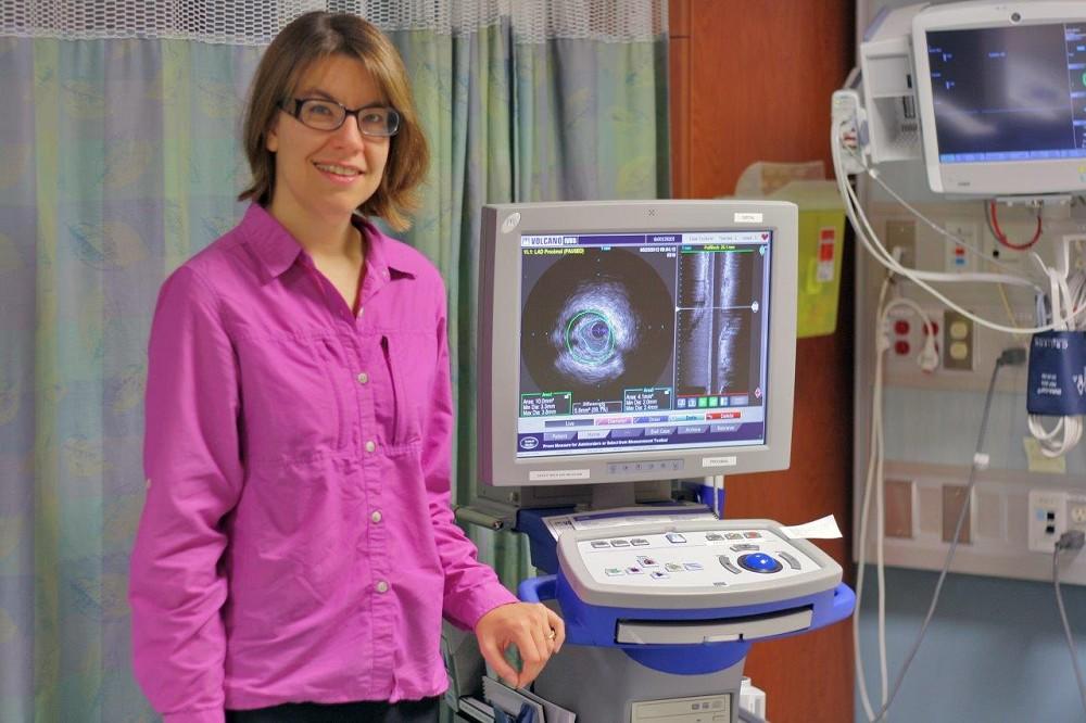 Dr. Andrea MacDougall, one of four interventional cardiologists at the Health Sciences Centre, shows how Intravascular Ultrasound (IVUS) gives a “close-up view” of the artery to determine the amount of plaque build-up along the artery walls.
A procedure used in the Catheterization (Cath) Lab at the Thunder Bay Regional Health Sciences Centre is helping interventional cardiologists like Dr. Andrea MacDougall assess blockages in coronary arteries more accurately.
Called “Intravascular Ultrasound” or IVUS, it is an ultrasound probe that is inserted to the artery like a regular angioplasty catheter to provide an image from within the artery. Available at the Health Sciences Centre since spring 2012, this equipment is proving to be an extremely useful tool in the Cath Lab for improving patient care.
Angiography shows two-dimensional images of the tube-shaped coronary arteries. In most cases, this is enough to identify tight blockages that might benefit from stenting or bypass grafting. There are, however, situations where two-dimensional images don’t give cardiologists all of the information they need.
“Sometimes when you get a narrowing in the artery it’s hard to tell how tight it is from a 2D image, especially if the plaque is asymmetrical,” Dr. MacDougall said. “IVUS allows us to do ultrasound from inside the artery. We can replay the images and take measurements, and get objective evidence about how tight an artery is.”
Cardiologists also use IVUS to visualize exactly where plaque is located. This can help to determine whether stenting is possible, or whether a patient might require surgery. IVUS can also be used to assess stents that have already been inserted, and to determine whether they are expanded properly.
Dr. MacDougall said that IVUS is a tool used with the angiogram, and would never replace it. The angiogram gives a “big picture” overview of the coronary arteries, which is usually enough to identify blockages. It is only in certain cases where there is a question about a particular spot that the IVUS helps, giving a “close-up” view of the artery. Dr. MacDougall said that she uses the IVUS in only about 1 in 12 cases.
But in those cases when it is used, it can have a huge impact on patient care. Dr. MacDougall described one of several cases in which the IVUS gave a more precise picture of a blockage, saving the patient travel time and possibly surgery.
“Just viewing the angiogram, I probably would have sent him off for bypass surgery,” Dr. MacDougall said. “Using the IVUS though, we saw that the narrowing wasn’t as tight as we thought, so we were able to treat him here in Thunder Bay with angioplasty.”
It also works the other way, where the IVUS has revealed serious blockages.
“Sometimes we find that the vessels are tighter than we think,” Dr. MacDougall said.
She mentioned one younger patient who had chest pains, though the reason wasn’t clear. All of her arteries looked normal except for one that seemed to have a slight taper in the left main artery – the one that supplies most of the heart with blood.
“We thought we’d investigate further using the IVUS, and found that it was actually 70-80% narrowed. It was a lot more serious in the ultrasound images than the angiogram led us to believe. The patient ended up going for a three-vessel bypass,” she said.
“IVUS has certainly helped my decision making,” Dr. MacDougall said.
IVUS was made possible thanks to your generous donations to the Northern Cardiac Fund. To support other potentially life-saving programs and equipment like IVUS, please visit our website at www.healthsciencesfoundation.ca or call us directly at (807) 345-4673. Thank you!