‘Game-changer’ in molecular imaging within sight
by Julio Heleno Gomes
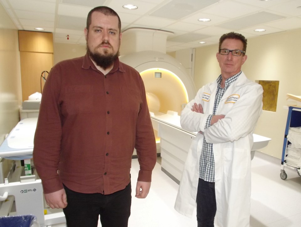 Dr. Mitchell Albert has been working on a medical imaging technique that uses xenon in MRIs. Here, Albert (R) is shown in front of an MRI scanner with Dr. Yurii Shepelytskyi (L).
(Via Tbnewswatch.com and Lakehead University’s Research Matters forum)
A technique being refined by researchers in Thunder Bay that provides razor sharp images of the lungs and blood flow images of the brain could revolutionize the future of personalized medicine, leading to earlier and more precise detection of different types of cancers and brain-related diseases.
The approach, referred to as hyperpolarized (HP) xenon-129 molecular imaging, promises more detail and sensitivity than other imaging techniques and does it instantaneously.
“It’s potentially a very powerful technique,” says Dr. Mitchell Albert, a Lakehead University professor in chemistry and Thunder Bay Regional Health Research Institute (TBRHRI) research chair in molecular imaging and advanced diagnostics.
Albert’s primary research is in the use of xenon in magnetic resonance imaging (MRI) technology. As a graduate student in the 1990s, he co-invented a technique to boost the MRI signal using HP xenon gas, for which he received several awards, including a U.S. Presidential Award from Bill Clinton.
He then spent 15 years at Harvard Medical School, improving the use of HP xenon as a diagnostic tool to detect small airway diseases such as COPD (chronic obstructive pulmonary disease), asthma, and cystic fibrosis. In 2011 he came to Thunder Bay, where he continues to work on these innovative imaging technologies.
This specific area of research – the imaging of blood flow in tissue, a process called “perfusion” – involves a patient inhaling xenon, a colourless, odourless gas used as a contrast agent for the imaging of soft tissues. The xenon is specially prepared in a polarizer to boost the MRI signal and is dispensed in a bag. With the subject holding their breath, the MRI monitors how the xenon dissolves and moves through blood vessels.
This technique was used to examine patients who suffered from “long COVID”, with symptoms ranging from breathlessness to “brain fog,” including headaches and dizziness. Utilizing xenon MRI, researchers were able to determine that not enough oxygen was getting into the red blood cells and, from there, to other organs.
“The job of the lungs is to deliver oxygen to the body, so people that had COVID had an impairment of that process,” Albert explains.
“That means they have a deficiency of getting oxygen into the bloodstream. That’s why they have poor ventilation. That’s why they were breathless. That’s why they had fatigue – their muscles, their cells weren’t getting enough oxygen. So we were able to get clues using our technique to help solve the mystery.”
At the same time, Albert and his team at TBRHRI started doing brain imaging, since that’s the organ with the highest blood flow. Focusing on people with Alzheimer’s disease, xenon imaging revealed that these patients had lower cerebral blood flow, as well as indicating atrophy or shrinkage of the grey matter. As a result, Albert’s team has proposed a biomarker that could monitor these types of diseases.
“We were able to make perfusion images of the brain, which is very important in all sorts of diseases of the brain, such as Alzheimer’s, Parkinson’s, stroke and brain tumours,” Albert says.
A member of Albert’s research team, Dr. Yurii Shepelytskyi, has helped conceptualize a xenon biosensor for molecular imaging. A native of Ukraine, Shepelytskyi studied applied physics at the national university in Kyiv, with a focus on microwave electrodynamics. While still in Kyiv, Shepelytskyi attended a lecture by Dr. Alla Reznik, a Thunder Bay-based researcher developing technology to spot early-stage breast cancer. This intrigued him to check out the research taking place in the city.
“I looked up the research conducted by Dr. Albert’s group, and its novelty and tremendous potential impact piqued my interest immediately,” he says.
Shepelytskyi moved to Thunder Bay and three years ago, obtained a PhD in chemistry and material sciences. He continued his research as a post-doctoral fellow and was recently appointed an adjunct professor in Chemistry. His work has involved programming the clinical MRI scanner, developing a mathematical model for xenon perfusion imaging, performing image reconstruction, data analysis and statistical analysis of the acquired data.
Currently, he continues developing and optimizing the performance of novel contrast agents for molecular MRI imaging as well as developing passive frequency-selective inserts for MRI that can substantially improve the quality and resolution of the acquired images.
“Hyperpolarized xenon-129 MRI paves the way for modern MRI into the realms of functional and molecular imaging,” Shepelytskyi says, explaining that identifying a specific type of cancer non-invasively reduces the amount of false-positive diagnoses and cuts down on the need for biopsies.
“This gives hyperpolarized xenon-129 MRI a unique opportunity to become a potential pillar for future personalized medicine and early-stage disease detection. Future development in this field can revolutionize modern health care and diagnostics,” he says.
In simple terms, the technique increases the xenon signal by several factors of magnitude. And by directing xenon in and out of the centre of large molecules, called “supermodular cages,” they could offer more detailed images of metabolic processes as they’re occurring.
“At the molecular imaging level the technique is so sensitive that we can see very, very small tumours and early-stage metastases wherever they go in the body,” Albert states.
“This xenon molecular imaging has the sensitivity of positron emission tomography or PET. PET is very sensitive but PET doesn’t have the good spatial resolution or localization. We’re doing it with MRI, which has excellent spatial resolution and anatomical localization. It only takes a fraction of a second to make the images, meaning we can catch things developing in real time.
“So it offers the best of both worlds: it has very, very high sensitivity and at the same time high spatial resolution. It’s also fast. All these elements are very important.”
Much of this work has been supported by grants from, among others, the Natural Sciences and Engineering Research Council of Canada (NSERC), the Ontario Research Foundation, the Ministry of Health, and Mitacs.
This molecular imaging technology is still at the experimental stage and is now in pre-clinical testing, Albert says, adding that his team is also collaborating with specialists in respirology and neurology for testing on patients with different ailments.
“It’s a game-changer for imaging lung function and brain function, absolutely, and has the potential to revolutionize molecular imaging.”
Research Matters highlights the important work of researchers at Lakehead University.