PET Molecular Imaging Laboratory Works Towards Cutting-Edge Diagnosis of Parkinson’s Disease
by Marcello Bernardo
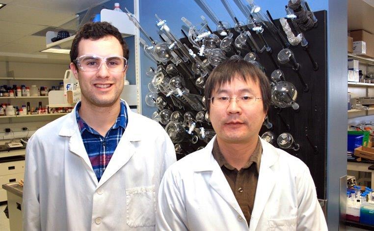 From L-R: Daniel Tesolin and Dr. Shusheng Wang.
PET molecular imaging laboratory works towards cutting-edge diagnosis of Parkinson’s Disease.
From L-R: Daniel Tesolin and Dr. Shusheng Wang.
PET molecular imaging laboratory works towards cutting-edge diagnosis of Parkinson’s Disease.As featured in the 2015/16 Thunder Bay Regional Research Institute Annual Report
Currently, there are no definitive tests or confirmed biomarkers that can be used to diagnose Parkinson’s disease. However dozens of research teams around the world are studying Glucocebrosidase (GCase) enzyme levels in the brain, which are lower in people with Parkinson’s disease. A few of those research groups – including scientists at the Thunder Bay Regional Research Institute’s Molecular Imaging Lab – are using advanced positron-emission tomography (PET) imaging to detect the disease earlier and more accurately. This research aligns with our first goal of creating Impact through Excellence in Imaging.
“We hope to create a way to detect Parkinson’s disease early so that we can slow its progression and treat its symptoms earlier,” said Daniel Tesolin, a Master’s degree student who returned to Thunder Bay from the University of Ottawa to work with Dr. Chris Phenix at the Research Institute.
GCase activity seems to drop rapidly at the onset of early Parkinson’s disease and leads to a build up of neurodegenerative plaques or Lewy bodies that damage brain cells and cause the symptoms of Parkinson’s disease.
“By the time Parkinson’s disease is diagnosed, typically 60% of the cells in the areas of the brain that control motor function have already died,” Tesolin said.
Tesolin and Dr. Phenix have spent the last year developing radiolabelled probes that will bind to the GCase and “light up” during a PET scan.
“PET has the advantage of being super sensitive,” Tesolin said. “With PET imaging, you can easily attach a radioactive isotope to a potential imaging compound without changing its biological function. The amount of isotope is so microscopic there are virtually no side effects.”
In 2015, the team developed a new molecule that has the potential to show GCase during PET imaging. The next step is to radiolabel their probe with fluorine 18 (18F), a man-made radioactive isotope created using the new cyclotron in Thunder Bay. Dr. Shusheng Wang joined the team in early 2016, applying his chemistry expertise to the probe development.
“We modify the structures of the potential probe molecules to develop ones that have the best properties for a diagnostic PET imaging agent. If we find that we have good results, then we will use the cyclotron facility for radiolabelling (and then biological testing),” Dr. Wang said.
Tesolin said that his return to Thunder Bay and this research would not be happening at the Research Institute without our new cyclotron. “We would have to ship our 18F from Hamilton or Winnipeg, but that’s not efficient because it has a half-life of 110 minutes,” he said. “We need the cyclotron onsite for quick access to 18F.”
The next step is to do pre-clinical research with the radioactive PET tracers which, if successful, could lead to human clinical trials. Such techniques could also be used to unravel the molecular mechanisms of other diseases.