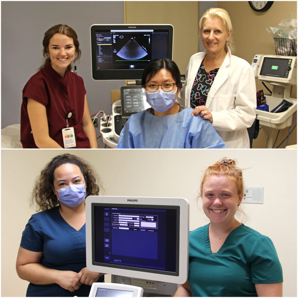
Celebrating Sonography Week (October 2-6)
by Marcello Bernardo
 Join us in thanking the sonography professionals at our Hospital this week for everything they do.
Sonography Week (October 2-6) is an opportunity to recognize and promote the profession of Diagnostic Medical Sonography (ultrasound) which plays a vital role in the care and treatment of community members.
Sonographers are medical detectives. They use their ultrasound training, technical skills, and understanding of the human body and its systems to decide if structures are normal or abnormal and adapt their investigation as they find clues throughout an examination. This information is then used by doctors to determine the necessary treatment or next steps for the patient.
A sonographer uses an instrument called a transducer or probe on a patient over the area of the body under investigation. The probe emits high-frequency sound which is inaudible to the human ear. As the probe is moved around it records echoes as sound waves bounce back to the ultrasound machine to determine size, shape and consistency of soft tissues. This information is relayed in real-time to produce images on a computer screen.
The quality of an ultrasound exam is very dependent on the skills of the sonographer who completed the scan. If they are not a great detective who takes in all the evidence and finds all the clues, then it is difficult to solve the case. As well, no two cases are ever the same, so a sonographer’s day is never dull.
Sonography is a growing, dynamic profession and sonographers are in demand in hospitals, medical imaging clinics and tertiary healthcare facilities. Many sonographers are also employed as educators, application specialists or sales representatives with medical equipment manufacturing firms, or as researchers.
Did you know?
- The gooey gel that people relate to ultrasound exams is necessary for sound waves to travel between the probe and the patient.
- Ultrasound exams can last between 20 and 60 minutes with the sonographer in very close proximity to the patient.
- Not all sonographers perform all types of exams. Learning different body systems requires special training and separate written and clinical examinations. Students graduate with knowledge of basic exams and can then further their specialization and knowledge if they desire.
- At TBRHSC, pregnancy ultrasound is a very small portion of our workload. Most exams at our site relate to abdomen, pelvic, arterial and venous, breast, scrotal, prostate, thyroid, neonatal brain and muscle and tendon studies.
The sonography programs in Canada vary in focus and length. Individuals can be trained in three areas: generalist, cardiac or vascular sonography. For more information and a list of accredited training programs, visit the Sonography Canada website: https://sonographycanada.ca/
Most sonographers LOVE ultrasound and love talking about it, so next time you come across one of these allied health professionals, ask them about their dynamic career.