What is Molecular Imaging?
by Marcello Bernardo
Imagine you and a dozen friends are meeting at a restaurant for dinner one evening. You all have smartphones with a special app that uses the GPS to show you where your friends are at any given moment. You’re the first to arrive, so you open the app to see all their blue dots moving towards you. Sally and John are on the highway. Jake is moving north on Memorial. Linda is on Water St. and just about to turn right onto Red River Road.
But Trevor, Cass, Richard, and Megan are all stopped at the corner of John St. and Algoma. In fact, they don’t move for a while. As the others arrive, you all look at this app wondering what has happened until finally they start moving again. A few minutes later they join you and tell you that there was an accident at that corner. Nobody was hurt, but traffic was stopped until the accident could be cleared up enough to move around.
This is kind of how molecular imaging works. Scientists develop special bits of material called probes that are injected into the body. These probes are similar to the GPS beacon in a smartphone except that instead of an app, you view these probes using diagnostic imaging. If everything is running as it should, these probes move throughout the body easily. If there is a problem like a cancer tumour or a blockage in an artery, the probes slow down or stop at that area – much as your friends did when there was an accident at the corner.
Molecular imaging is already being used at the Thunder Bay Regional Health Sciences Centre. For example, PET/CT imaging is used as a minimally invasive way to diagnose cancer. Using a nuclear tracer, doctors can locate tumours and determine the cancer’s stage. It also helps during chemotherapy treatment to determine if the tumours are shrinking. Molecular imaging is also being used in many other ways including finding and assessing blockages in arteries to diagnose low blood flow to the heart.
Perhaps most exciting is the fact that TBRRI scientists are working on brand new methods of imaging. One example is new technology using hyper-polarized gas that is 100,000 times more specific to assess conditions like asthma and COPD.
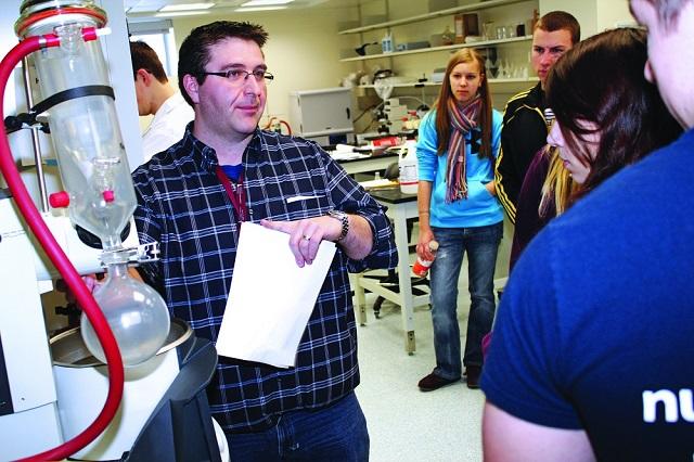
There are several different kinds of molecular imaging probes. Scientists at the TBRRI are developing many new ones right now, including for breast cancer patients. Using molecular imaging, doctors can find diseased areas, design personalized treatments for patients, and monitor how effective those treatments are for a clearer picture of your overall health.
Thank you to all the people and organizations who have supported the TBRRI through the Thunder Bay Regional Health Sciences Foundation to make it the world class research centre it is today! You along with our government and industry partners have helped our scientists develop new probes for diagnostic imaging – and there are many more to come. Support local research by making your donation to the Health Sciences Discovery Fund online (healthsciencesfoundation.ca) or by calling the Foundation at (807) 345-4673. Together, we’re building a healthier, wealthier, and smarter Northwestern Ontario.